Live webinar ASI op 23 oktober 2019 door Dr. Ron Hochstenbach

ASI October newsletter: What are the advantages of Light Microscopy in Cytogenetics and Pathology?
Light Microscopy has the ability to detect and visualize the most sensitive of signals and abnormalities in both cytogenetic and pathology diagnostics.
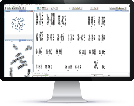
With an automated Metaphase Finder, the best chromosomes can be karyotyped for the analysis of numerical or structural changes, thus enabling the identification of rare genetic diseases as well as the pathogenesis of malignancies. See HiBand for more details.
In IHC cells are automatically segmented while in FISH signal detection is categorized for greater statistical results. This expedites the workflow process plus compares thousands of cells with the original H&E sample. See HiPath Pro for more details.
Mark your Calendar: October 23rd
Back by popular demand, we are proud to present a live webinar on WGS and Light Microscopy by Dr. Ron Hochstenbach. Click here to sign up.


